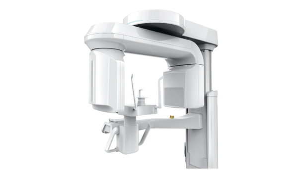
Conebeam CT scanner

Why do you need a 3D x-ray?
Traditionally, dental surgeons were using 2D x-rays on film to see all or a specific part of the dentition, intraosseous structures and jaws of a patient. Several years ago, the advent of 3D digital radiography has greatly facilitated the work of dental surgeons, in addition to reducing the dose of radiation for the patient. The most widely used 3D radiography in implantology is panoramic radiography that shows teeth and bone structures of the maxillofacial area.
To schedule the installation of dental implants, it is often necessary to obtain additional information which is unfortunately not available with traditional 2D x-rays:
- Quantity and density of the alveolar bone;
- Position and anatomy of the maxillary sinus (for implants in the upper jaw);
- Precise position of the inferior alveolar nerve (for the implants in the lower jaw).
These three elements must absolutely be considered due to complications that can occur during the implant surgery. Fortunately, in recent years, digital 3D radiography, also called Cone Beam Computed Tomography (CBCT), made its appearance in some dental offices and its use is becoming more widespread.
What does it require?
Major investments are often made by dental surgeons to obtain devices that can take digital x-rays in 3D, but the advantages of using this technology are largely worth the cost.
The difference between 2D and 3D x-rays
3D x-ray devices used for the study of maxillofacial structures are equipped with a fixed-anode x-ray tube just like 2D devices.
The main difference is that in CBCT 3D units, the scanner emits a conical x-ray beam, while in traditional 2D x-ray machines, it is a triangular-shaped beam.
A lower dose of x-rays
The CBCT 3D scanner rotates around the patient’s head while emitting a low x-ray dose compared to that emitted by traditional 2D x-ray devices.
The images captured by the scanner can be viewed as a two-dimensional or a three-dimensional model.

A single 3D x-ray can generate hundreds of images
The scanning software collects the data that allow producing different views. This same software also provides access to a multitude of features that the dental surgeon can use to plan implant surgery. A single three-dimensional x-ray can generate hundreds of images and even 3D videos of the patient’s face.
3D X-rays for sharper images
The quality of images obtained by computed tomography (3D x-rays) is significantly greater than traditional 2D x-rays. The main innovations derived from 3D scans are the lack of distortion and high resolution obtained by the powerful data reconstruction software. A single acquisition of data by a CBCT allows creating multiple views that can be manipulated in all directions by the dental surgeon.
Benefits of 3D digital X-rays
Three-dimensional x-rays offer several advantages for the dental surgeon and the patient.
For the patient:
- Less exposure to radiation.
- Easier to understand the explanations of the dental surgeon (clarity and accuracy of radiographic images). Multiple views can be displayed, enabling the dental surgeon and the patient to see the structures from different angles.
For the dentist:
- The quality and quantity of information provided by the CBCT on different types of tissues and organs (soft tissues, bones, muscles, nerves and blood vessels), facilitates the insertion of implants, making it more precise, quick and predictable. The dental surgeon can get pictures of the impacted teeth, measure the quality and volume of the jawbone, sinuses, and inferior alveolar nerve, which are not available with traditional 2D x-rays.
- The dental surgeon knows the ideal position for each implant, with millimeter precision, which facilitates the surgery and allows him to achieve unmatched accuracy.
Damage to surrounding structures (remaining teeth, inferior alveolar nerve, and maxillary sinus) can be minimized.


